FAQ Library
Practical guides on AI-assisted radiology — how the models work, what they actually detect, where they fall short, and how they fit into daily dental workflows.
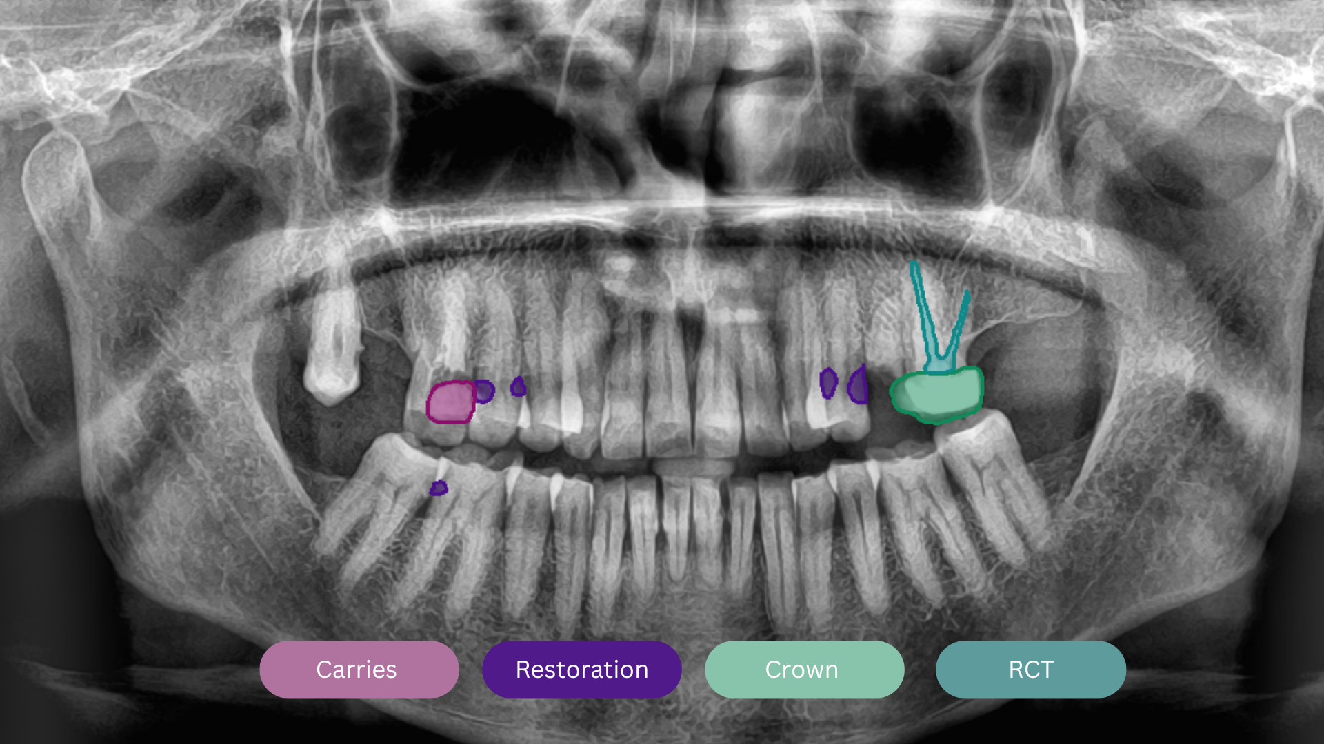
Scans an OPG in under 30 seconds. Flags caries, bone loss, periapical lesions, and impactions — with per-finding confidence scores the dentist can review before signing off.
Explore X-Ray AI →Showing 15 of 15 articles
01
Deep learning segments early, moderate, and advanced lesions across all tooth zones — each with a per-tooth confidence score the clinician can accept, override, or flag.
02
From image preprocessing and zone segmentation to multi-label detection and structured report output — this is the pipeline behind every scan, explained plainly.
03
How the model tells apart periapical granulomas, abscesses, and cysts — on both panoramic and periapical views — and where it still struggles with overlapping anatomy.
04
Obturation length, fill density, and apical seal — the AI evaluates all three and flags under-filled or over-extended cases for the treating dentist to review.
05
Beyond detection — the model tracks bone-to-implant contact, peri-implant bone levels, and crestal bone loss over time. Useful for long-term follow-up, not just diagnosis.
06
CEJ-to-crest measurement, horizontal vs. vertical defect classification, and AAP/EFP Stage I–IV staging — from a single scan, without manual caliper work.
07
Impaction detection, angulation classification, and proximity-to-IAN flagging on panoramic X-rays. Particularly useful for third molar extraction planning and risk conversations.
08
Mucosal thickening, fluid levels, and maxillary sinus opacification — the kind of incidental findings that prompt ENT referrals and, sometimes, save patients a second scan.
09
Sensitivity, specificity, and AUC benchmarks — including head-to-head comparisons with general dentists and specialists. The numbers are encouraging, but the caveats matter too.
10
Image quality degradation, rare pathologies, overlapping anatomy — known failure modes, explained honestly. AI doesn't replace clinical judgment, and this article doesn't pretend otherwise.
11
The evidence-based answer is no — with nuance. This covers when AI augments specialist interpretation well, when it doesn't, and why full automation is still premature in most clinical contexts.
12
Per-tooth analysis covering caries depth staging, periapical bone changes, PDL space widening, and post/crown/core detection — the same output format as OPG, applied to periapicals.
13
Where volumetric AI stands right now — what's commercially available, what's still in research, and what Medecro's roadmap looks like. Honest about the gap between OPG and CBCT capability.
14
Condylar flattening, erosion, osteophytes, and asymmetry — detectable on panoramic radiographs and clinically relevant far more often than most practitioners realize.
15
Upload → AI analysis → dentist review → patient report → share. With real time benchmarks from clinics currently on the platform and notes on where it connects to your existing CMS.